
Scientists developed a microscope that fits in a needle to get a realtime look inside the human
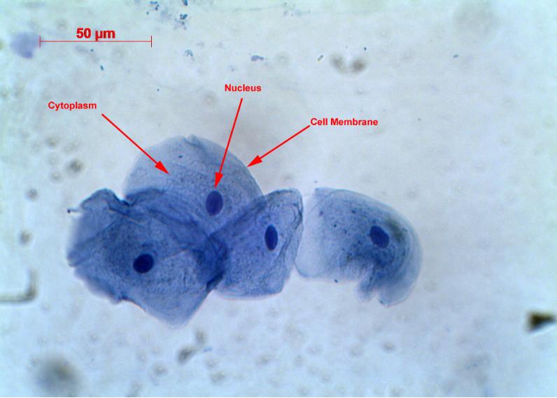
Remove any excess solution by allowing a paper towel to touch one side of the coverslip. Place the slide on the microscope, with 4 x or 10 x objective in position and find a cell. Then view at higher magnification. Methylene blue stains negatively charged molecules in the cell, including DNA and RNA. This dye is toxic when ingested and it.
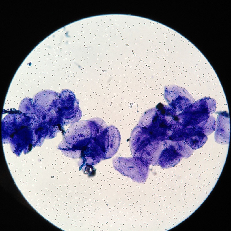
Some cells under the microscope. 大鲵小站
In Figure 3.1.2 3.1. 2, only one edge of the tissue slice has epithelial cells. In Figure 3.1.2 3.1. 2 A that edge is indicated with an arrow, but when looking at a specimen under a microscope, you have to figure out for yourself where the edge with the epithelial cells is. Figure 3.1.2 3.1. 2: A slice of a trachea.
Stunning Microscopic View of Human Skin Cells Wins 2017 Nikon Small World Competition News
How does the human body look like under an electron microscope?Objects Under Electron Microscope! (Part - 1). Watch Here https://youtu.be/c6Jqis6wrbkMusic: h.

Cheek Cell Lab Aiden's Blog
Observing human cheek cells under a microscope is a simple way to quickly view and learn about human cell structure.
Are we really made up of microscopic cells? conspiracy
The Human Cheek Cell. 1. List the 3 parts of the Cell Theory. 2. Describe or define each of the following. Draw your cells to scale. 4. Why is methylene blue necessary? 5. The light microscope used in the lab is not powerful enough to view other organelles in the cheek cell.. We also acknowledge previous National Science Foundation.
Cell Under Electron Microscope Video Bokep Ngentot
Mitosis in an animal cell. Cells from the Chinese Hamster Ovary are shown undergoing mitosis. Beginning with a cell spread on the substrate, follow prophase,.

blood cells, cells, human, electron microscope, scan, blood, microscopic, medicine, microbiology
A Guide to Microscopic Structure of Cells, Tissues and Organs Robert L. Sorenson Table of ConTenTs ChapTer 1 InTroduCTIon and Cell ChapTer 2 epIThelIum ChapTer 3 ConneCTIve TIssue ChapTer 4 musCle TIssue ChapTer 5 CarTIlage and bone ChapTer 6 nerve TIssue ChapTer 7 perIpheral blood ChapTer 8 hemaTopoesIs ChapTer 9 CardIovasCular sysTem
February 2011 Cell As a Unit of Life
Cheek Cells Under a Microscope Requirements, Preparation and Staining Cheek cells are eukaryotic cells (cells that contain a nucleus and other organelles within enclosed in a membrane) that are easily shed from the mouth lining. It's therefore easy to obtain them for observation. Some of the main parts of a cell include: 1.

10,151 Human Cell Under Microscope Images, Stock Photos & Vectors Shutterstock
Browse 1,185 authentic human cells under microscope stock videos, stock footage, and video clips available in a variety of formats and sizes to fit your needs, or explore cancer cells or dna stock videos to discover the perfect clip for your project. Browse Getty Images' premium collection of high-quality, authentic Human Cells Under Microscope.
Cells Rumney Marsh Academy Science Revere, Massachusetts
The Human Body Under the Microscope | Discover Magazine The Sciences Mind Technology Health Environment Planet Earth Lifestyle / Planet Earth The Human Body Under the Microscope A visual voyage through the cells, organs, microbes and molecules that make up our bodies. By Colin Salter Feb 11, 2015 10:00 AMNov 18, 2019 4:30 PM Newsletter

Human Cells Under Microscope HighRes Stock Photo Getty Images
Observerving cells under a microscope Microscopy, size and magnification.. Even larger human cells - like the skin cell - are 20 times smaller than a grain of salt. A red blood cell is much.

Full HD. Many living dividing cells under microscope, magnification 400X Stock Footage AD ,
hemoglobin iron red blood cells microscope Human blood contains many different components, from white blood cells to platelets, but the most abundant component by far are red blood cells. More properly known as erythrocytes, red blood cells make up 70% of an adult human's cells by count.
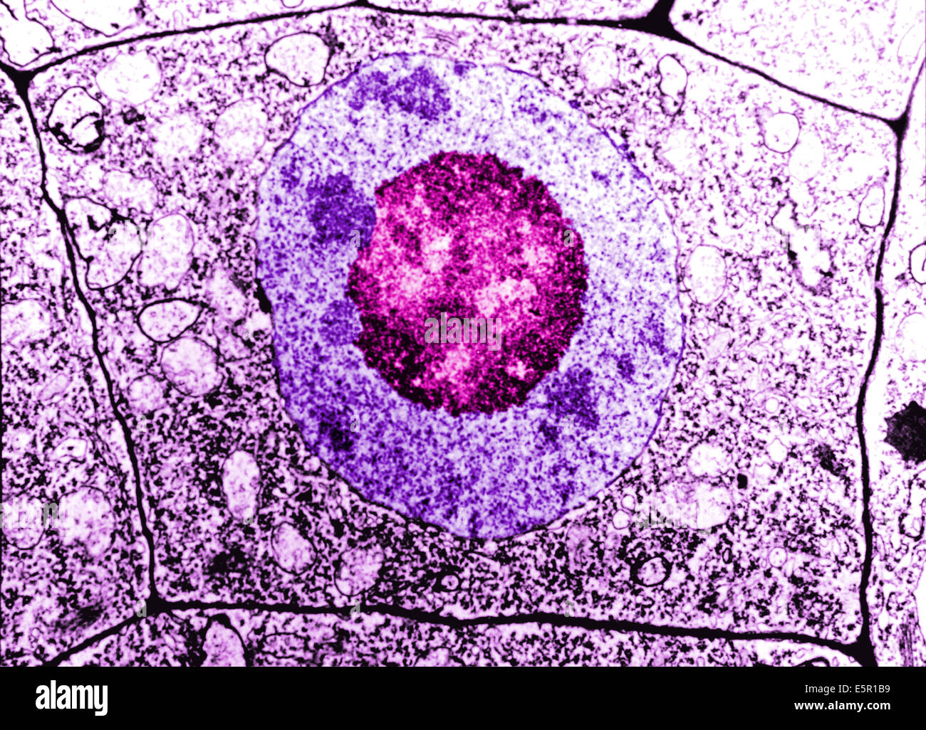
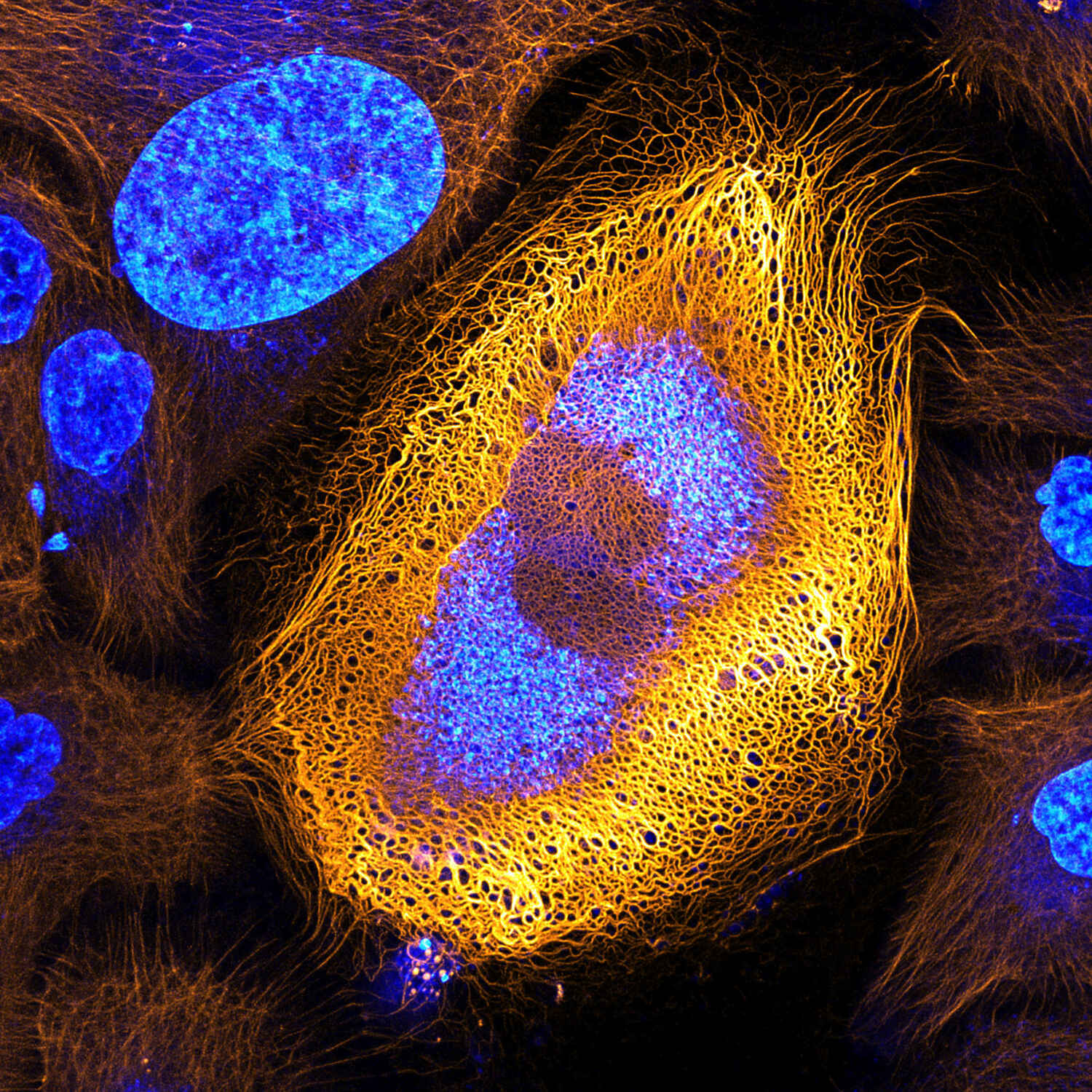
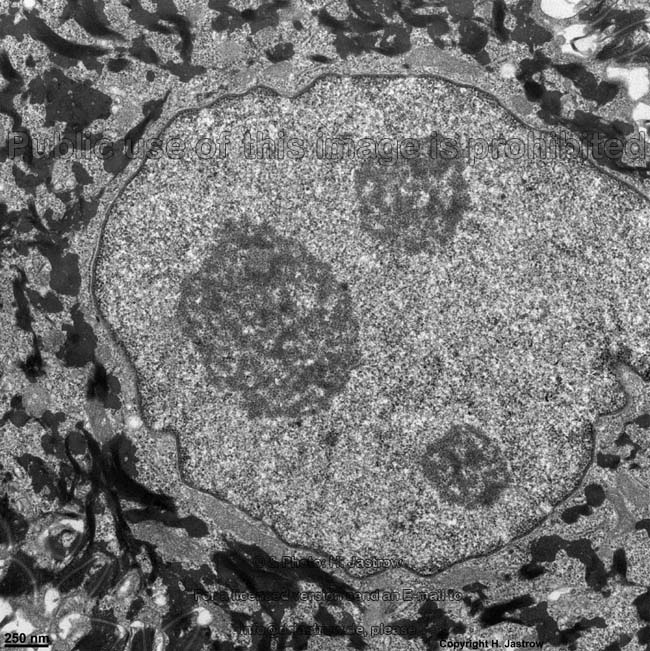
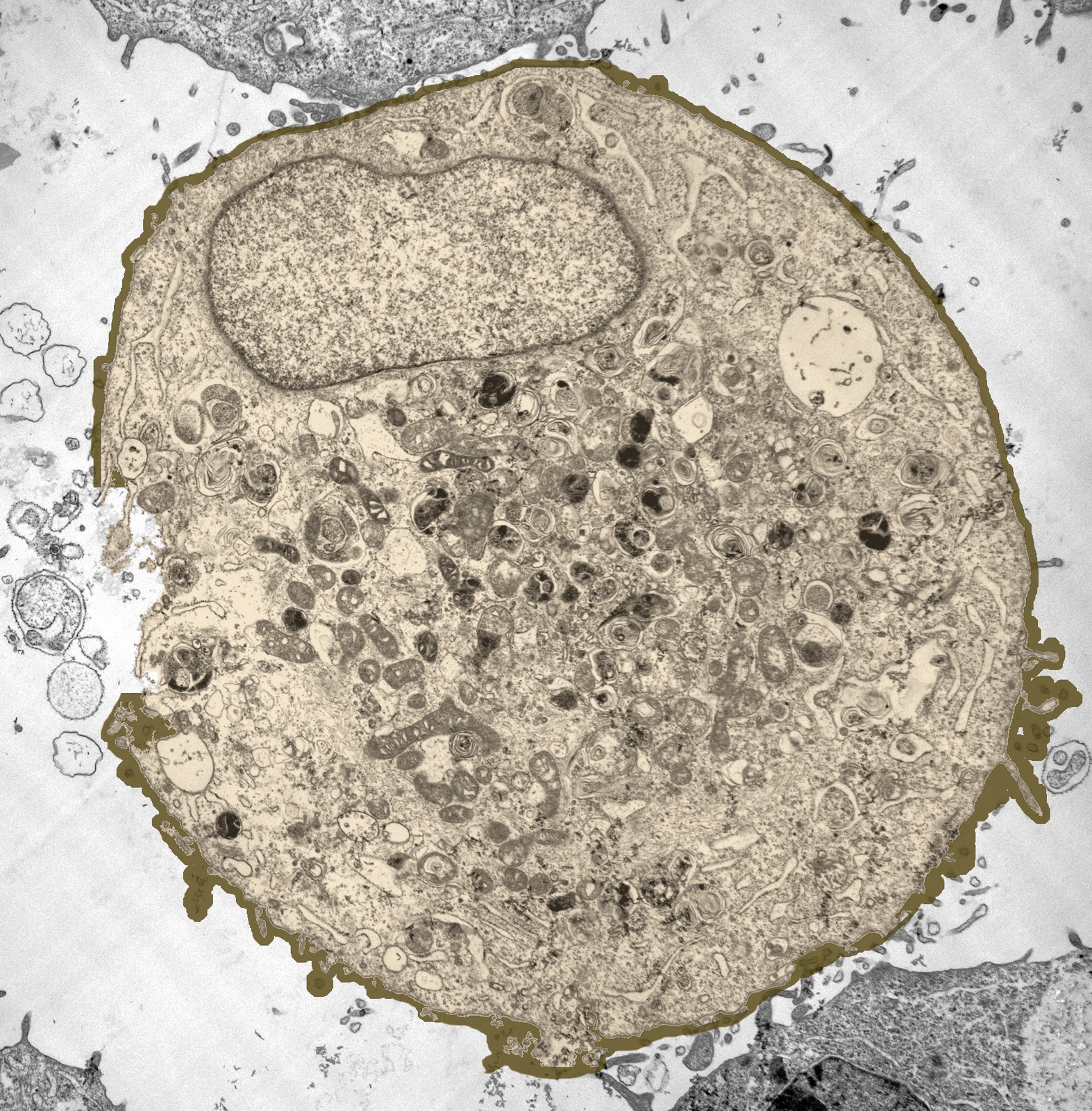
Electron Microscopy of a normal human cell, The cell membrane, nucleus and the nucleolus are all
Human cheek cells are made of simple squamous epithelial cells, which are flat cells with a round visible nucleus that cover the inside lining of the cheek.C.

And these are our lung cells. Microscopic photography, Things under a microscope, Biology art
Human Cheek Cells Figure 3. Human cheek cell at 400x zoom.. View under the microscope using the highest magnification for the best cellular details and draw what you see. Be sure to indicate the magnification used and specimen name. Also, indicate the estimated cell size in micrometers under your drawing. Figure 7.
4.2 Discovery of Cells and Cell Theory Human Biology
Science Is Beautiful: The Human Body Under The Microscope A new book showcases the magnificent micro-world By Lydia Ramsey | Published Feb 3, 2015 10:34 PM EST Science Science Is Beautiful, a new.

Researchers Identify Protein that Makes Skin Cancer Cells More Invasive Onco'Zine
Cells and Tissues. Tissues are classified into four basic types: epithelium, connective tissue (includes cartilage, bone and blood), muscle, and nervous tissue. Organs are assembled from the four basic types of tissues and have cells with specialized functions. These slides contain tissue sections that are easily confused with each other.